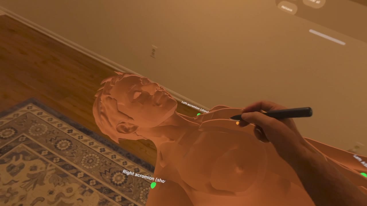
I mark a few points on the body
A stylus or a pinch tags anatomical landmarks such as the acromion, xiphoid process and hip peaks. Each point turns green as it is confirmed.
Augmented-reality trauma ultrasound
Intelliview uses patented AI and augmented reality to map a patient's internal anatomy from a handful of points on the skin, then guides anyone to a specialist-grade trauma ultrasound, right at the point of injury.
Actual headset capture · Intelliview early prototype on Apple Vision Pro
The problem
A soldier caught in a blast. A driver pulled from a wreck on a rural highway. On the outside, they may look fine. On the inside, they're bleeding. In a hospital, an ultrasound specialist would find it in minutes.
The technology
Our patented technology, developed in conjunction with the Johns Hopkins Applied Physics Laboratory, predicts where a patient's organs are from just a handful of points on the outside of the body, then shows that map on the patient through augmented-reality glasses.
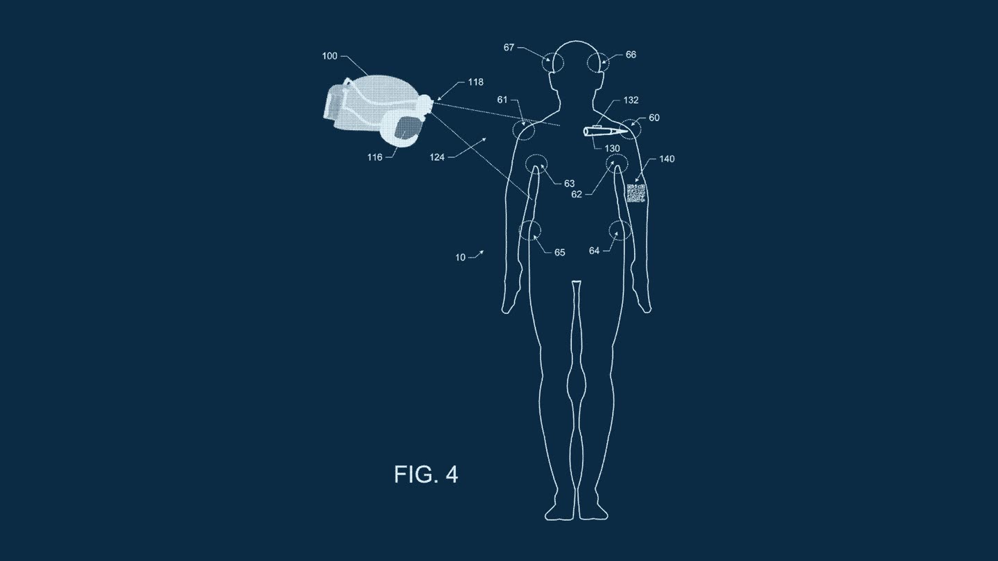
The user touches a handful of external points on the patient: shoulders, sternum, hips. No CT scanner, no imaging suite.
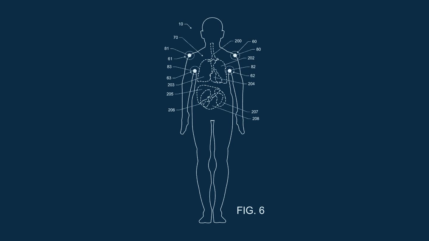
A model trained on hundreds of CT scans predicts the size and position of 66 structures in the chest and abdomen for this specific patient.
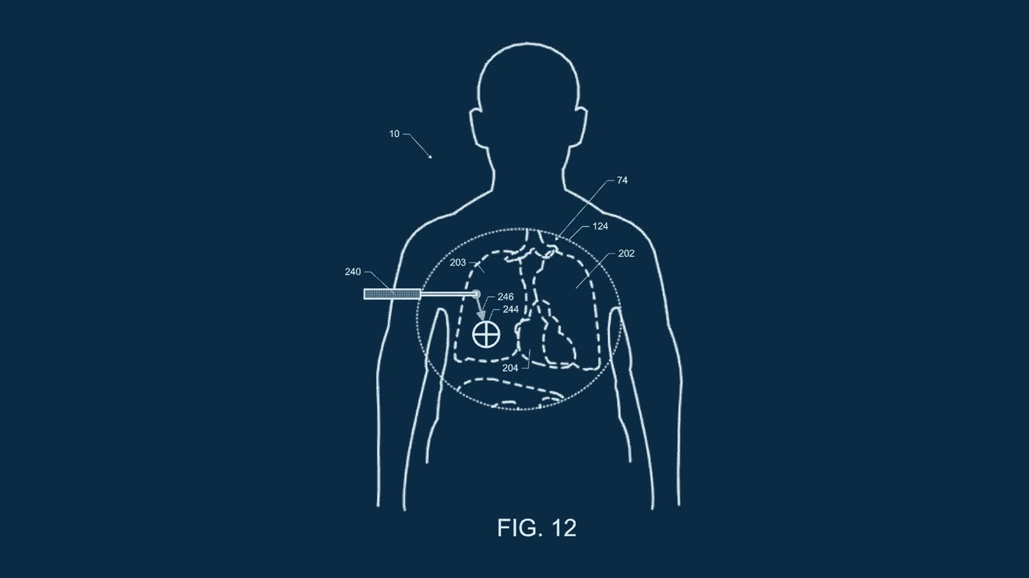
The predicted anatomy appears on the patient, and guides a portable ultrasound probe to exactly the right spot for each view of the exam.
In early studies, people with no medical training at all used the system to perform an eFAST exam.
The prototype
The original research ran on HoloLens. We are rebuilding it as a product, starting with an early prototype on Apple Vision Pro. Every image below is captured from the headset.
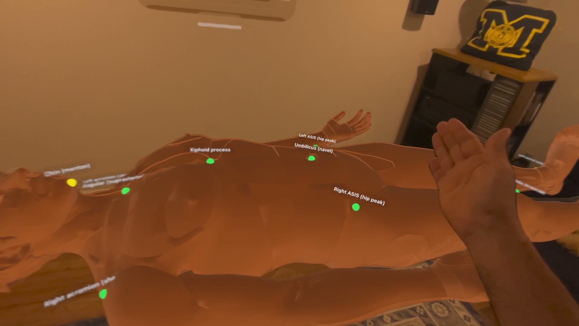
A stylus or a pinch tags anatomical landmarks such as the acromion, xiphoid process and hip peaks. Each point turns green as it is confirmed.
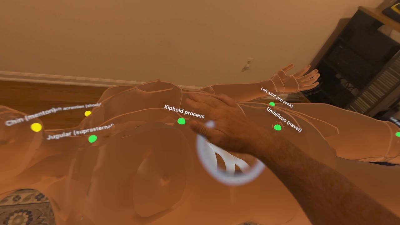
Registration scales and aligns a full anatomical model to the captured landmarks, so what you see lines up with the body in front of you.
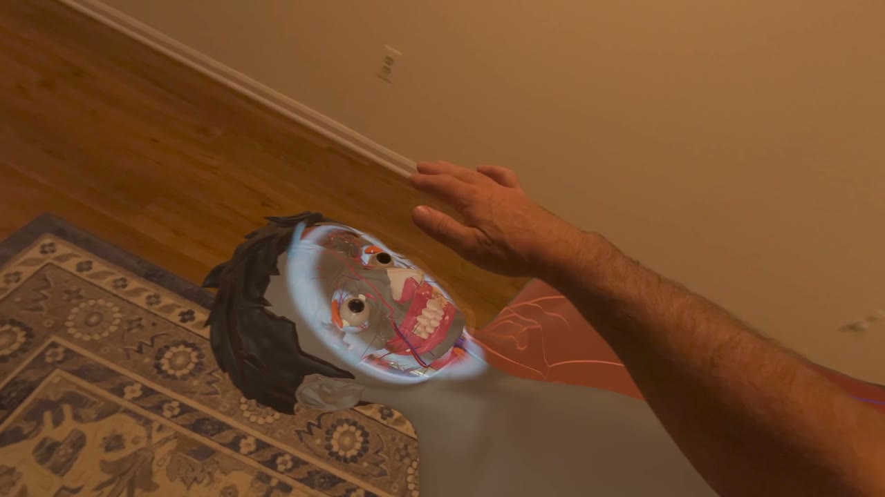
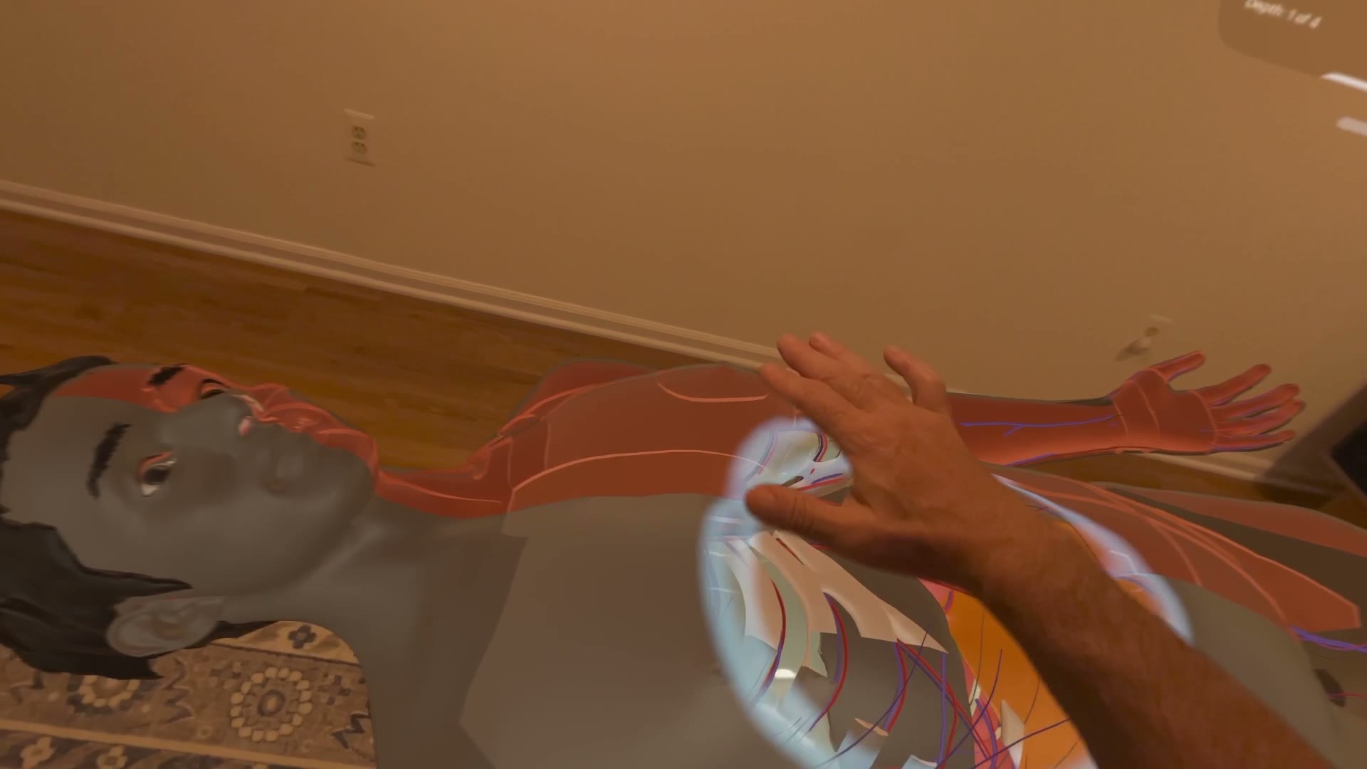
An open hand works like a scanner, dissolving the surface to reveal what lies beneath, right where it is.
Connect the probe so the live image appears exactly where it touches the body.
A physician hundreds of miles away can stand beside the medic and guide the scan.
Markets
Point-of-injury assessment, where the nearest radiologist may be hours away.
Know what's bleeding before the patient reaches the hospital.
Triage many casualties when specialists are scarce.
Specialist-grade imaging without a specialist on site.
Team

Chief Executive Officer · Co-founder
Interventional radiologist with more than two decades of reading the scans Intelliview teaches anyone to take.
LinkedIn ↗
Chief Technology Officer · Co-founder
Thirty years turning government research into shipping products, across medical devices, government R&D and enterprise software. EE, University of Michigan. Builder of Intelliview's Vision Pro prototype.
LinkedIn ↗We're looking for clinical, defense and investment partners to take it the rest of the way.
Get in touch